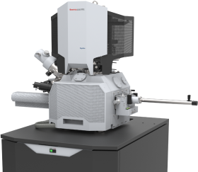
Aquilos 2 Cryo FIB-SEM

The Aquilos 2 is a state-of-the-art Focused Ion Beam Scanning Electron Microscope (FIB-SEM) designed specifically for the precise and site-specific milling of cellular samples, optimized for cryo-EM tomography. It features an integrated fluorescence light microscope (iFLM), allowing for accurate targeting and visualization of fluorescently labeled regions, enhancing both precision and efficiency in sample preparation.

We are equipped to mill both waffle grids and standard plunge-frozen grids, enhancing flexibility and compatibility with various cryo-EM sample preparation methods.

Instrument Parameters


  • Electron Optics
  • Gun Type
    High-stability Schottky field emission gun
  • Resolution (with cryo-stage)
    1.6 nm at 30 kV, 2.6 nm at 2 kV (at room temperature), 6.0 nm at 2kV (at cryo-temperature)
  • Beam Current
    1.5 pA to 400 nA
  • Accelerating Voltage Range
    200 V – 30 kV
  • Ion Optics
  • Column Type
    Sidewinder ion column
  • Resolution (with cryo-stage)
    7.0 nm at 30 kV
  • Beam Current Range
    1.5 pA – 65 nA
  • Accelerating Voltage Range
    500 V – 30 kV
  • Detection System
  • In-lens segmented lower (T1) and upper (T2) detectors
  • Everhart-Thornley SE detector (ETD)
  • Integrated Nav-Cam™ camera for navigation
  • IR camera for chamber observation
  • Cryo-Stage and Cryo-System
  • Temperature
    Operates at < -170°C for vitrified sample preservation
  • Stage Movement
    XY Range: 110 mm, Z Range: 65 mm, Tilt Range: -15° Tilt range at cryo (eucentric WD): -15° to +55°, Rotation: 360° (endless)
  • Cooldown Time
    Less than 20 minutes
  • Cryo-stage chamber vacuum
    <8e–5 Pa.
  • Cryo-Loader
    Load-lock system supporting sample transfer with integrated shutter protection
  • Integrated Hardware
  • Sputter Coater
    In-chamber, retractable, for platinum deposition, prevents charge buildup during imaging
  • Gas Injection System (GIS)
    Provides protective coatings during milling
  • iFLM Correlative System (Integrated Fluorescence Light Microscope)
  • System
    Widefield optical microscope
  • Objective Lens
    20x Zeiss Epiplan-Apochromat, NA 0.7 (Piezo-driven)
  • Working Distance
    1.3 mm
  • Imaging Field of View
    500 × 500 µm (700 µm diagonal)
  • Filters
    Semrock LED-DA/FI/TR/Cy5-B-000 Quadband filter set for fluorescence channel separation
  • LED source
    CoolLED system with four wavelengths (365, 450, 550, 635 nm)
  • Camera
    Basler ace 2 (2A4504-5gmPRO; Sony IMX541 CMOS sensor)
  • EasyLift NanoManipulator for Cryo-Lift-Out
  • Cryo-Lift-Out
    Enables precise removal and transfer of site-specific regions from high-pressure frozen samples to cryo TEM grids
  • Temperature Control
    Maintains -165°C at the needle tip to ensure sample integrity during lift-out
  • Software
  • xT Software
    Comprehensive control for cryo-sample preparation and lamella milling
  • AutoTEM Cryo Software
    Automated lamella preparation and batch processing
  • Maps Software
    Correlation of light microscopy and SEM data; supports up to 64k x 64k imaging datasets